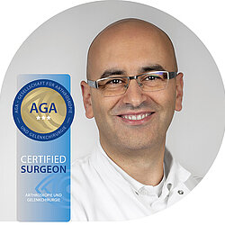
Center for Special Joint and Trauma Surgery


Whether sports injury, accidental injury, fracture of the upper or lower extremity, consequence of injury or wear-related disease - our team of experts at the Center for Special Joint and Trauma Surgery will be happy to advise you on injuries or diseases of the shoulder, elbow, knee or ankle joint. After careful analysis of your complaints, we will draw up a treatment concept tailored to your individual needs. The most modern surgical and treatment methods are used, taking into account the latest scientific findings. Thanks to the extensive experience of our specialists, you benefit from the highest level of routine and expertise.

If surgery becomes necessary, we use minimally invasive or arthroscopic procedures. Most joint injuries or diseases can be treated extremely precisely by arthroscopy. As a patient, you benefit not only from the smaller skin incision, but also from less pain, faster rehabilitation and a better functional result due to less scarring as a result of the tissue-sparing procedure.

Our primary goal is the preservation of the joint or the restoration of joint function. We can also offer joint-preserving procedures for incipient osteoarthritis.

If joint preservation is no longer possible, bone-saving endoprostheses that can be individually adapted to you are available.

Treatment focus

Knee

  • meniscus suture, meniscus transplantation
  • all cartilage therapy procedures including cartilage cell transplantation
  • anterior cruciate ligament reconstruction (hamstring, quadriceps or patellar tendon graft)
  • posterior cruciate ligament reconstruction
  • cruciate ligament revision surgery
  • complex ligament stabilization (multi-ligament injuries)
  • Leg axis correction
  • stabilization of the kneecap (MPFL-plasty, trochleaplasty, tuberosity offset)
  • surgical stabilization of tibial plateau fractures, patella fractures and joint fractures of the femur

Shoulder

  • Extension of the acromion and removal of the calcific deposit
  • Capsule release for frozen shoulder
  • Rotator cuff suture
  • Capsular reconstruction for mass defects of the rotator cuff
  • Shoulder stabilization including bony glenoid reconstruction
  • Treatment of long biceps tendon disorders
  • Stabilization of the acromioclavicular joint
  • surgical therapy of early shoulder arthrosis (CAM procedure)
  • insertion of an artificial shoulder joint (anatomical and inverse total shoulder endoprosthesis)
  • Shoulder total endoprosthesis replacement surgery
  • treatment of glenoid fractures of the shoulder
  • surgical stabilization of fractures of the upper arm and clavicle

Elbow

  • Surgical therapy of tennis and golfer's elbow
  • Stabilization of the unstable elbow including ligament reconstruction (LUCL, MUCL)
  • Cartilage therapy of the elbow
  • capsule release for elbow stiffness
  • Surgical therapy of early elbow osteoarthritis
  • Insertion of an artificial elbow joint and radial head prosthesis
  • nerve decompression at the elbow (Sulcus ulnaris syndrome)
  • surgical stabilization of joint fractures of the upper and lower arm

Disease patterns knee

Leg axis malposition (bow or knock-kneed)

Women tend to have knock-knees, while men usually have bow legs. The deviation of the leg axis is not pathological per se, but it is a risk factor for the development and progression of cartilage damage. For example, bow-leg malalignment leads to an increased load on the inside of the knee, while bow-leg bending leads to an increased pressure load on the outside of the knee. The more pronounced the malalignment, the more pronounced the incorrect loading. As a result of the leg malposition, cartilage and meniscus damage occurs in the respective knee section, which can lead to arthrosis in the long term. If the leg axis is corrected in time, the development of arthrosis can be prevented and the need for an artificial joint replacement can be avoided or delayed.

Conservative measures (special insoles and orthoses) can reduce the increased pressure on the joint section, but they cannot correct the leg axis.

Surgical correction of the leg axis can eliminate the incorrect loading. Before surgery, an exact analysis of the leg skeleton must be performed. The surgical intervention is performed in combination with a mirror examination of the knee joint, during which any meniscus and cartilage damage is treated. Depending on the malposition, the correction of the leg axis is performed through small skin incisions on the upper or lower leg. Rarely, correction is necessary on both the upper and lower leg.

After the operation, the leg must not be fully loaded for six weeks.

Posterior cruciate ligament

The posterior cruciate ligament is one of the central knee stabilizers. A tear of the posterior cruciate ligament occurs much less frequently than an injury to the anterior cruciate ligament. The leading symptom is pain, rather than a feeling of instability. Isolated injuries of the posterior cruciate ligament can be treated conservatively. However, combination injuries with tearing of the knee joint capsule or the collateral ligaments are frequently present. In these cases, posterior cruciate ligament surgery with reconstruction of the capsule and collateral ligament structures is necessary.

Posterior cruciate ligament reconstruction is performed as a short inpatient procedure (two to three days). The follow-up treatment after posterior cruciate ligament reconstruction is more complex than after anterior cruciate ligament reconstruction. A special knee orthosis must be worn for eight weeks. Relief of the operated leg on forearm crutches is necessary for four weeks, and sports (soccer, handball, etc.) can only be practiced again after nine months.

Patella

There is pain and recurrent dislocation of the patella in the presence of patellar misalignment (patella) or patellar instability. Failure of conservative treatment requires an accurate root cause analysis (patella height, trochlear dysplasia, TTTG distance, torsion determination, capsular ligament laxity, leg axis analysis) in order to perform targeted surgical therapy. The most common treatment options include:

  • lateral retinaculum lengthening
  • MPFL plasty
  • Tuberosity offset
  • Trochleaplasty

After arthroscopic assessment and treatment of any cartilage damage, the above. operations are performed through small skin incisions to eliminate the misalignment of the patella and restore patella stability.

Depending on the surgical procedure, partial weight-bearing on the operated leg is required for between two and six weeks.

Cartilage damage

Cartilage damage to the knee joint can result from: direct trauma, wear and tear, cruciate ligament injuries, meniscus damage, leg axis misalignment, or patellar deformity.

Minor cartilage damage usually causes little discomfort, but can progress and eventually lead to osteoarthritis. The cartilage itself has little potential for regeneration. The treatment of cartilage damage is therefore the domain of surgical therapy.

Surgical treatment must be preceded by a precise analysis of the causes and concomitant pathologies in order to achieve a good long-term surgical result. This includes the testing of knee stability as well as the examination of the leg axis.

During arthroscopy, the extent, depth and localization of the cartilage damage are determined and the most suitable treatment procedure is selected. Bone marrow stimulating procedures (nanofracturing), matrix-induced procedures (AMIC), cartilage-bone transplantation (OATS) or autologous cartilage cell transplantation (ACT) are used. Crucial for the success of cartilage therapy is not only to select the appropriate cartilage procedure, but also to recognize concomitant pathologies and to treat them as well. This requires additional interventions such as cruciate ligamentoplasty, meniscus therapy or leg axis correction.

Cartilage therapy can be performed arthroscopically or through a small skin incision. After the operation, you must not put full weight on the operated leg for six weeks. It takes three to six months before you can resume sporting activities.

Cruciate ligament revision surgery

Cartilage damage to the knee joint can result from: direct trauma, wear and tear, cruciate ligament injuries, meniscus damage, leg axis misalignment, or patellar deformity.

Minor cartilage damage usually causes little discomfort, but can progress and eventually lead to osteoarthritis. The cartilage itself has little potential for regeneration. The treatment of cartilage damage is therefore the domain of surgical therapy.

Surgical treatment must be preceded by a precise analysis of the causes and concomitant pathologies in order to achieve a good long-term surgical result. This includes the testing of knee stability as well as the examination of the leg axis.

During arthroscopy, the extent, depth and localization of the cartilage damage are determined and the most suitable treatment procedure is selected. Bone marrow stimulating procedures (nanofracturing), matrix-induced procedures (AMIC), cartilage-bone transplantation (OATS) or autologous cartilage cell transplantation (ACT) are used. Crucial for the success of cartilage therapy is not only to select the appropriate cartilage procedure, but also to recognize concomitant pathologies and to treat them as well. This requires additional interventions such as cruciate ligamentoplasty, meniscus therapy or leg axis correction.

Cartilage therapy can be performed arthroscopically or through a small skin incision. After the operation, you must not put full weight on the operated leg for six weeks. It takes three to six months before you can resume sporting activities.

Meniscus

Damage to the medial or lateral meniscus can result from changes in wear and tear or accidents (twisting of the knee), as well as being a consequence of knee joint instability (cruciate ligament injury). Meniscal tears rarely heal with conservative treatment. Tears of the meniscal base (meniscal suture) or suspension (root refixation) should be reattached to restore the shock absorbing function of the meniscus. For other types of tears, partial removal of the meniscus is necessary. In these cases, as much as necessary but as little meniscal tissue as possible should be removed to avoid unduly compromising meniscal function. If little meniscal tissue can be preserved, it may be necessary to sew in a meniscal implant (CMI) or transplant a meniscus (meniscal allograft).

Meniscus procedures are performed arthroscopically and usually on an outpatient basis. After a partial meniscus removal, you can bear full weight. After a meniscus suture, partial weight-bearing is necessary for between two and four weeks, depending on the localization, as well as wearing a knee orthosis.

Tibial plateau fracture

A tibial plateau fracture is a serious injury to the joint-forming tibial plateau caused by an accident. The injury usually requires surgical treatment. A computed tomography scan is required to accurately assess the extent of the fracture and to plan surgery.

Simple fracture forms such as bony avulsion injuries of the cruciate ligament or a submerged joint surface can be treated and stabilized arthroscopically. For complex fracture forms, an open procedure is necessary. Depending on the location of the fracture, a skin incision is necessary on the outer and/or inner side of the knee joint, and possibly even in the popliteal fossa. The fracture is set up open and stabilized with screws and special metal plates.

Since the tibial plateau heals slowly, follow-up treatment is lengthy. The load-bearing capacity of the operated leg is only given after 10 weeks in the case of complex fractures.

Anterior cruciate ligament

The anterior cruciate ligament is one of the central stabilizers of the knee joint. A tear of the anterior cruciate ligament is one of the most common ligament injuries of the knee joint and usually results in joint instability with recurrent bending of the leg. Due to the instability, there is a high risk of secondary injuries (meniscus and cartilage damage).

A tear of the anterior cruciate ligament usually does not heal stably, so that patients with high functional demands should be treated surgically. During arthroscopy, the torn cruciate ligament is replaced by a tendon (graft) produced by the patient's own body. The femoral flexor tendons (hamstring tendons), the patellar tendon and the femoral extensor tendon (quadriceps tendon) are used here. The choice of graft is based on your personal needs and requirements.

The procedure is performed on a short inpatient basis (two to three days). After an anterior cruciate ligament reconstruction, you can resume full weight-bearing after three weeks at the latest, loose running or cycling are possible after three months, stop-and-go sports (soccer, handball, etc.) after nine months.

Disease patterns shoulder and elbow

Osteoarthritis of the elbow

Like any other joint, the elbow is subject to wear and tear changes, which occur in particular after bone fractures, elbow dislocations/instabilities or rheumatic diseases. The wear and tear leads to pain and increasing restriction of movement.

If conservative arthrosis therapy does not bring the desired success, a joint-preserving intervention with arthroscopic arthrolysis (see elbow stiffness) can be performed for milder forms of arthrosis. In severe forms of arthrosis with joint destruction, joint-preserving surgery is not promising. In these cases, an artificial joint must be inserted. In this case, the destroyed joint surfaces are removed through a skin incision on the back of the elbow and an artificial joint adapted to the individual anatomy is inserted.

An inpatient stay for seven days is required. The operated arm is immobilized for a short time. Movement exercises must be performed daily and weight bearing must be avoided for six weeks.

Arthrosis of the shoulder joint

The most common causes of osteoarthritis of the shoulder joint include changes in wear and tear (aging process), consequences of accidents involving fractures of the humeral head, and late effects of rotator cuff damage (rotator cuff defect arthropathy).

Regardless of the cause of the osteoarthritis, there is destruction of the articular cartilage at the humeral head and glenoid cavity. This results in an inflammatory reaction with pain and increasing restriction of movement of the affected shoulder joint.

If conservative treatment has not led to the desired success, surgical intervention must be considered. If limitation of movement is the primary concern, joint-preserving surgery with removal of excess bone attachments and release of the thickened joint capsule can be performed. If pain is the main concern, an artificial joint replacement should be considered.


Is an artificial shoulder joint an option for you?

You will benefit from an artificial shoulder joint if the following complaints are present:

  • severe pain that interferes with your everyday life (e.g. when dressing, combing your hair or performing personal hygiene)
  • Pain at rest, which especially disturbs your night's rest
  • Restriction of shoulder mobility and loss of strength
  • Persistent complaints despite exhaustion of conservative treatment options (anti-inflammatory and analgesic drugs, cortisone injections, physiotherapy)

Various shoulder endoprosthesis models are available, from which we will select the most suitable model for you:

  • Total joint replacement (anatomic total shoulder arthroplasty):
    The prerequisite for an anatomic total shoulder arthroplasty is an intact rotator cuff. In an anatomic joint replacement, the destroyed joint surfaces are removed. The humeral head is replaced with a metal hemisphere and the glenoid cavity with a plastic socket. In recent years, bone-saving stemless shoulder prostheses have increasingly replaced the larger stem-anchored prostheses, as this allows the individual anatomy and thus the shoulder function to be restored even more accurately.
  • Reverse shoulder joint replacement (inverse total shoulder arthroplasty):
    Reverse joint replacement is used in cases of large, irreparable rotator cuff defects (rotator cuff defect arthropathy), non-reconstructible fractures of the humeral head or endoprosthesis replacement surgery.
    In reverse joint replacement, the ball and socket are interchanged. That is, the metal ball is attached to the scapula, and the socket is attached to the humeral head. With the help of the hooded muscle, the arm can then be raised or spread apart again, even with a defective rotator cuff.
  • Endoprosthesis replacement surgery:
    Although artificial shoulder joints (total shoulder arthroplasties) function just as reliably as artificial hip or knee joints, shoulder arthroplasty failure can occur, as with other artificial joints. This is most often due to loosening, infection or a secondary defect in the rotator cuff. In such a case, replacement of the artificial shoulder joint may be necessary.

After artificial shoulder joint replacement, immobilization of the arm is necessary. During the day, however, the operated arm can be used as early as the second postoperative day, for example for feeding. Since the artificial joint must grow into the bone, rehabilitation measures are not advisable until the sixth postoperative week.

Elbow luxation / elbow instability

The elbow is the second most dislocated joint after the shoulder. Depending on the severity of the injury, dislocation can result in rupture of the lateral collateral ligament (LUCL), the capsule, the medial collateral ligament (MUCL) and/or fractures of the coronoid process and the radial head. If only ligamentous structures are injured and the elbow is stable after dislocation, the injury can be treated conservatively in a humeral splint. More extensive injuries or additional fractures should be treated surgically.

Chronic ligament injuries of the elbow do not necessarily manifest as instability, but rather as decreased weight-bearing capacity and pain.

Clinical examination leads the way in acute or chronic elbow injury. Magnetic resonance imaging can be used to visualize the injured structures and identify the extent of the injury. In the case of bony injuries, a computer tomography may also be necessary.

An endoscopy of the elbow can confirm the diagnosis, especially in inconclusive cases. Depending on the extent of the injury, stabilization of the elbow is performed through one or two incisions, which are used to reattach the capsule and collateral ligaments to the bone, as well as stabilize any fractures.

In chronic injuries, the collateral ligaments may need to be reinforced with an endogenous tendon (e.g., gracilis or palmaris longus tendon).

The stabilization procedure is performed under inpatient conditions. The arm is immobilized in an upper arm splint for six weeks after the operation, and movement exercises must be performed daily.

Elbow stiffness / free joint bodies

After injuries, circulatory disorders of the bone or due to changes in wear and tear, free joint bodies may form and the movement of the elbow may be restricted.

Physical therapy can treat movement restrictions that exist over a short period of time well. If there are free joint bodies with recurrent entrapment or if there is a prolonged high-grade restriction of elbow motion, surgery is indicated.

The procedure can be performed arthroscopically. The inflamed joint mucosa, the free joint bodies, excessive bony attachments and the severing of the scarred joint capsule are removed. In the case of severe movement restrictions, the ulnar nerve (sulcus ulnaris syndrome) must also be detached.

The procedure can be performed on an outpatient basis. Intensive physiotherapy over six weeks is necessary.

Elbow tunnel syndrome (Sulcus ulnaris syndrome)

After injuries, bone fractures, in the context of inflammatory diseases or due to movement restrictions of the elbow, irritation of the ulnar nerve with pain and sensory disturbances in the ring and little finger can occur.

If conservative treatment does not lead to the desired result or the neurologist can determine a higher degree of damage to the ulnar nerve, surgery is indicated.

Through a small skin incision on the inside of the elbow, the ulnar nerve is accessed and scarring is released. Depending on the pathology, the nerve is moved out of the bony bed to the flexor side.

The procedure can be performed on an outpatient basis.

Fracture of the ulnar process (olecranon fracture)

A fracture of the olecranon process is a common injury from falling on the elbow. Undisplaced fractures can be treated conservatively.

Due to the pull of the triceps muscle on the fractured olecranon process, there is a high risk of the fracture ends coming apart. Surgical setting up of the fracture is indicated if the fracture ends have moved apart. The fracture is reduced via a skin incision on the extensor side and stabilized by a plate with screws.

Immobilization is only necessary for the first few days, and weight-bearing is possible again after six weeks.

Golfer's elbow (Epicondylitis humeri ulnaris)

Golfer's elbow is the counterpart to tennis elbow. Prolonged overuse of the wrist and finger flexor muscles causes painful inflammation of the tendon insertions at the inner humeral cartilage of the elbow.

The condition responds well to conservative treatment (cooling, physiotherapy with stretching and detonation of the muscles, epicondylitis orthosis).

Only if conservative treatment is unsuccessful can surgery be considered. During surgery, tendon attachments of the flexor muscles are released and inflamed tendon tissue is removed. The procedure is performed through a small incision on the elbow.

The treatment can be performed on an outpatient basis. The operated arm should be rested for six weeks.

Calcareous shoulder

Calcified deposits in the rotator cuff cause distension of the tendon with secondary bursitis and shoulder tenderness. In most cases, these are smaller calcific deposits that can be treated well conservatively.

However, conservative treatment often fails for large calcific deposits. In these cases, arthroscopy is performed. The distended tendon attachment is superficially slit in the area of the calcific deposit before the calcific deposit can be cleared out. Removal of the inflamed bursa is obligatory, and bony expansion of the acromion may also be required.

Immobilization of the operated arm is not required. The operated arm should be rested for six weeks. The follow-up treatment after calcific deposit evacuation is somewhat more protracted, so that complaints may persist for up to six months after the operation.

distal humerus fracture (supracondylar humerus fracture)

Fractures of the humerus distal to the body (supracondylar humerus fractures) occur particularly in children. Undisplaced or slightly displaced fractures can be treated conservatively. Displaced fractures require surgical stabilization. Childhood fractures can usually be reduced in a closed fashion. For stabilization, small wire pins are inserted through mini-incisions in the inner and outer humeral cartilages to stabilize the fracture.

The humerus must be immobilized in a splint for four weeks, and the wire pins can be removed after six weeks.

In adults, distal fractures of the humerus are usually displaced, requiring surgery. The surgery is performed through a skin incision on the extensor side. The fracture is set up open and usually stabilized by two plates. Depending on whether the articular surface is involved, it may be necessary to remove the olecranon process to visualize the joint fracture. The fracture can then be set up anatomically, stabilized with plates, and the olecranon process subsequently refixed.

After surgery, immobilization in an upper arm splint is required for four weeks. Daily range of motion exercises must be performed. Weight bearing can be done after six weeks at the earliest.

Immobilization of the operated arm is not required. The operated arm should be rested for six weeks. The follow-up treatment after calcium depot removal is somewhat more protracted, so that complaints may persist for up to six months after the operation.

Long biceps tendon

The long biceps tendon attaches over the upper cartilaginous joint lip (biceps tendon anchor, SLAP) at the glenoid top and runs through the top of the shoulder joint at the front. Tears of the long biceps tendon or injuries to the biceps tendon anchor (SLAP) can result from an accident, overuse, or changes in wear and tear, causing strain pain over the anterior shoulder segments.

Leading the way in finding a diagnosis is the clinical examination. Magnetic resonance imaging is helpful, but often cannot provide a definitive diagnosis.

The diagnosis can often only be made unequivocally with surgery. If the condition is still stable, smoothing of loose parts of the joint lip is sufficient. If the biceps tendon anchor is unstable, stabilization is necessary. Fresh lesions are reattached to the upper glenoid rim using special suture anchors (SLAP repair). In chronic degenerative lesions, however, reattachment does not produce satisfactory results. In chronic lesions, the biceps tendon is often not affected in isolation but is associated with other shoulder pathologies (rotator cuff lesion). In these cases, the long biceps tendon is simply detached at the superior glenoid rim (biceps stenotomy) or the tendon is anchored outside the joint at the humeral head (biceps stenodesis). Which of the above procedures is used depends on the pathology present as well as your personal requirements and wishes.

The operation is performed arthroscopically. Immobilization is only required for a SLAP repair.

Humeral head fracture / Bony avulsion of the rotator cuff

Bony avulsions of the rotator cuff (greater tuberosity fractures) are often the result of shoulder dislocation. If the fracture ends are not displaced, the injury can be treated conservatively by immobilization in a shoulder brace. Displaced fractures must be set up and stabilized surgically (arthroscopically or openly).

Fractures of the head of the humerus (subcapital humerus fractures) occur especially in older people with osteoporosis (bone loss). Undisplaced fractures can be treated conservatively by immobilization in a shoulder brace. Displaced humeral head fractures, on the other hand, should be treated surgically. The bone fractures are then set up open and stabilized with a plate or nail and screws. If the articular surface of the humeral head is shattered, it may not be possible to restore it, so the installation of an artificial shoulder joint may be necessary.

Rotator cuff

Accidents or changes in wear and tear can cause rotator cuff tendons to tear. A torn tendon does not grow back on its own.

Untreated tears increase in size over time, i.e. the tendon stumps retract and the associated muscles become irreversibly fatty. Defects that have existed for a longer period of time may then no longer be able to be closed.

Magnetic resonance imaging (MRI) shows the extent of rotator cuff damage and makes it possible to assess whether a tear has already existed for a longer period of time and whether the defect can still be closed surgically.

Conservative treatment may be considered only in cases of low functional demand, older patients, or defects that cannot be reconstructed.

Rotator cuff damage should be treated surgically. The procedure is performed arthroscopically. First, the tear configuration is determined, then the tendon stumps are mobilized before being reattached to the bone via special suture anchors.

If the tear exists over a longer period of time (years) and several tendons are affected, the defect may not be able to be completely closed. In this case, partial closure of the defect is performed to restore the best possible force closure between the individual muscles (margin convergence). Recently, a patch can also be inserted into the remaining defect, thus closing the joint to the acromion (superior capsular reconstruction).

The procedures are usually performed arthroscopically. Only in the case of very large defects is the tendon repaired via a small skin incision.

Collarbone fracture

The clavicle is the only bony connection of the shoulder to the rib cage. A clavicle fracture significantly impairs shoulder function. Most clavicle fractures can be treated conservatively (without surgery). The injury is then immobilized in a shoulder brace. Surgery is necessary when the broken bones are far apart or threaten to impale the skin. In such a case, the fracture is surgically set up and stabilized with a nail or plate with screws.

After surgery, immobilization of the shoulder is required for a maximum of four weeks. However, the arm can be used from the first postoperative day out of the bandage without weight bearing.

Shoulder blade fracture

Fractures of the scapula are rare injuries. Most of these fractures can be treated conservatively. Because the scapula is surrounded by strong muscles, fractures of the scapula usually heal without problems by immobilizing the arm in a shoulder brace. Only in cases of pronounced displaced fractures or involvement of the glenoid portions of the scapula (glenoid), surgical setting up of the fracture and stabilization may be required. Glenoid fractures in particular can be set up and stabilized arthroscopically.

Shoulder dachalgia (impingement syndrome)

Shoulder pain during abduction movements of the arm is often due to bursitis resulting from shoulder tendinitis. Treatment of shoulder tendinitis is the domain of conservative therapy with rotator cuff strengthening exercises and cortisone injections if necessary.

If conservative treatment fails, surgery may be necessary. In a procedure called subacromial decompression, the inflamed bursa is removed and the sliding space under the acromion is widened.

The procedure is performed arthroscopically. Immobilization of the arm is not necessary. Nevertheless, the operated arm should be rested for four weeks after the operation.

Acromioclavicular joint

The clavicle is the only bony structure that connects the shoulder to the trunk via the acromioclavicular joint (AC joint). Every movement with the arm exerts a great force on the acromioclavicular joint, which explains the susceptibility to the development of osteoarthritis. Pain is particularly present during overhead activities or when supporting oneself with the arm and is usually localized directly over the acromioclavicular joint.

Osteoarthritis of the acromioclavicular joint often responds poorly to conservative treatment because of a mechanical problem involving narrowing of the acromioclavicular joint space. During arthroscopy, the articular surfaces of the clavicle and acromion are smoothed and the acromioclavicular joint space is widened to restore proper joint space.

Immobilization is not required after surgery. However, the arm should be rested for six weeks postoperatively.

Shoulder instability / dislocation of the shoulder

The shoulder is the joint in the human body that dislocates most frequently. In the case of dislocation (shoulder luxation), typical damage occurs to the shoulder:

  • Tear of the capsule and the cartilaginous lip of the joint
  • rupture of the glenoid rim
  • indentation of the humeral head (Hill-Sachs dent)

Any shoulder dislocation should be reduced as soon as possible. Any damage that has occurred can be detected on X-ray and MRI (magnetic resonance imaging). Depending on the joint damage that has occurred, your age and your requirements, we will choose the most suitable therapy for you individually.

One-time dislocations may be treated conservatively. However, a recurrence of dislocation proves shoulder instability, so that surgery is then indicated.

During surgery, the torn cartilage lip (labrum) and capsule are reattached with special suture anchors (Bankart repair). Acetabular fractures sheared off with the labrum or capsule can also be reconstructed in this way (Bony-Bankart repair). Chronic bony acetabular rim defects are reconstructed by a bone chip from the iliac crest (Bone block) or stabilized by an offset of the coracoid process to the anterior inferior acetabular rim (Latarjet coracoid transfer). In large, centrally extending, hooking Hills-Sachs defects, it may also be necessary to suture the posterior joint capsule into the defect to prevent the Hill-Sachs dent from hooking again (remplissage).

The procedures can usually be performed arthroscopically. After surgery, immobilization of the shoulder is required for four weeks.

Frozen Shoulder

Frozen shoulder can develop after accidents, operations or spontaneously. The symptoms are pain, especially at rest, and an increasing restriction of shoulder movement. The disease leads to shrinkage, scarring and thickening of the joint capsule.

Frozen shoulder usually responds well to conservative treatment with physiotherapy and cortisone tablets.

If symptoms persist or there are contraindications to cortisone treatment, shoulder mobility can be restored surgically. This involves releasing the thickened and scarred joint capsule of the shoulder, thus restoring the mobility of the joint. The procedure is performed arthroscopically.

Immobilization is not required. After the operation, movement exercises must be performed consistently for the first few weeks.

Fracture of the radial head (radial head fracture)

Fractures of the radial head occur in the course of a fall and interception with the outstretched arm. The fractures also frequently occur in combination with elbow dislocation.

Undisplaced fractures can be treated conservatively by short-term immobilization in a humeral splint. Displaced fractures, on the other hand, should be treated surgically. Through a small skin incision on the outside of the elbow, open setup and stabilization with screws or a plate is performed for fractures of the radius neck. If the bone is shattered, the head of the radius usually cannot be reconstructed. In this case, implantation of a radial head prosthesis (radial head prosthesis) may be required.

After open fracture or implantation of a radial head prosthesis, only short-term immobilization of the arm is required. Weight bearing should be avoided for six weeks.

Stabilization of the acromioclavicular joint (AC stabilization)

The clavicle is the only bony structure that connects the shoulder to the trunk via the acromioclavicular joint (AC joint). A fall on the shoulder can cause the acromioclavicular joint (AC joint) to move apart with tearing of the ligaments between the clavicle and the coracoid process.

The diagnosis and extent of instability (Rockwood classification) is made from radiographs held. Mild injuries (Rockwood I+II) can be treated well conservatively. For more severe injuries (Rockwood III-V), surgical stabilization is indicated. Under arthroscopic view, a special suture system is inserted between the coracoid process and the clavicle, which reduces the acromioclavicular joint, allowing the torn ligaments to heal (AC repair). In the case of chronic injuries (accident more than three weeks past), the torn ligaments must also be replaced with an autologous tendon (for example, gracilis tendon) (AC reconstruction).

After the operation, the shoulder must be immobilized for six weeks. Weight bearing is possible after three months at the earliest.

Tennis elbow (epicondylitis humeri radialis)

Prolonged overloading of the extensor muscles of the wrist and fingers can

lead to painful inflammation of the tendon insertions at the outer humeral cartilage of the elbow.

The condition usually responds well to conservative treatment (cooling, physiotherapy with stretching and detonation of the muscles, epicondylitis orthosis).

Only if conservative treatment is unsuccessful can surgery be considered. During surgery, the tendon attachments of the extensor muscles are released and inflamed tendon tissue is removed. The procedure can be done arthroscopically or through a small incision.

The treatment can be performed on an outpatient basis. The operated arm should be rested for six weeks.

Our medical team


Dr. Markus Leyh

Senior Physician

Special Joint and Trauma Surgery

    Dr. Christian Ohm

    Senior Physician

    Special Joint and Trauma Surgery

      Elie Hassoun

      Senior Physician

      Special Joint and Trauma Surgery

        Contact & Appointment


        Dagmar Alms

        Secretariat Special Joint and Trauma Surgery

        If you have not found a date that suits you, please feel free to contact us by phone.

        Private outpatient clinic

        Phone +49 2351 945-2305
        Fax +49 2351 945-2307
        sekretariat.beck@hellersen.de

        Office hours

        0

        Tuesday
        8.00 - 15.00
        Appointments by appointment only

        Outpatient Clinic

        Phone +49 2351 945-2331
        Fax +49 2351 945-2258
        ambulanz@hellersen.de

        Office hours

        0

        Monday - Friday
        8.00 - 15.00
        Appointments by appointment only

        At all other times, you will be helped in our Central Emergency Outpatient Clinic Phone +49 2351 945-0.

        Central Emergency Outpatient Clinic